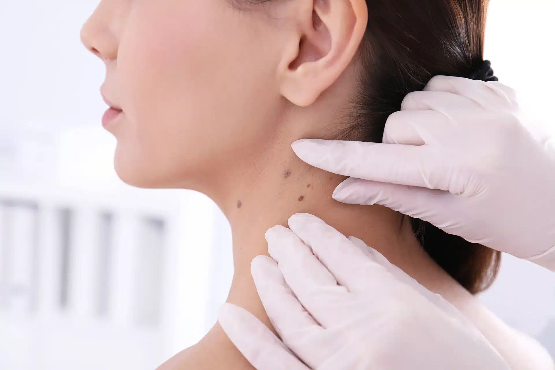
- Skin cancer is less common in Singapore, but it can still occur.
- Melanoma, basal cell, and squamous cell carcinoma are the three main types of skin cancer.
- Melanoma in Asian skin often appears on palms, soles, and nails.
- The ABCDE rule helps identify suspicious moles, but it is not diagnostic.
- Dermatoscopy provides information beyond a standard visual examination.
- Regular skin cancer screening may be appropriate for people with higher risk factors.
Skin cancer screening in Singapore plays an important role in evaluating suspicious skin changes before they become more advanced. While the overall risk of skin cancer is lower in Asian populations than in fair-skinned populations, it can still occur and may not always be recognised early.
For patients, the question is often straightforward: “Is this mole, spot, or skin growth something I should be concerned about?” Assessment by a dermatologist can help answer that question through a careful evaluation of your skin and individual risk factors.
Ahead, we will explain when skin cancer screening may be recommended and the warning signs that should not be ignored.
What Skin Cancer Screening Actually Is (and How It's Different from Mole Removal)
Many people assume that a skin cancer screening is simply a consultation to remove a suspicious mole. In reality, screening and mole removal serve different purposes.
A skin cancer screening is a medical assessment performed by a dermatologist to identify skin lesions that may require monitoring, further investigation, or treatment. The goal is to determine whether a mole, spot, or skin growth shows features that warrant closer attention.
The primary goal of a dermatologist’s mole check in Singapore is not to remove lesions immediately, but to examine your skin for signs that may be associated with skin cancer or other skin conditions. This often involves assessing not only the lesion that concerns you, but also the rest of your skin for additional findings that may otherwise go unnoticed.
A screening typically includes a visual examination of the skin and, when appropriate, the use of a dermatoscope. This handheld device provides magnification and specialised lighting, allowing the dermatologist to evaluate structures beneath the skin surface that are not visible to the naked eye.
If a lesion appears suspicious, further investigation may be recommended. This could involve monitoring the area over time or performing a biopsy, where a small sample of tissue is examined under a microscope to establish a definitive diagnosis.
Importantly, screening does not automatically lead to treatment. Many lesions assessed during skin cancer screening are ultimately found to be benign. The purpose of the examination is to distinguish lesions that can be safely observed from those that may require further evaluation.
If you’re considering the removal of a mole for cosmetic reasons, the process and goals are different from a medical skin cancer assessment.
Skin Cancer in Singapore: What the Local Picture Looks Like
When people think about skin cancer, they often picture fair-skinned populations in countries such as Australia, the United States, or parts of Europe. Skin cancer in Singapore is indeed less common than in many Western countries, largely because higher levels of melanin provide some natural protection against ultraviolet (UV) radiation.
However, lower risk does not mean no risk.
Dermatology literature has consistently shown that skin cancer can occur in Asian populations and may sometimes be diagnosed at a later stage. One reason is lower awareness. Many people do not consider themselves at risk and may delay seeking medical attention for a changing mole or unusual skin lesion.
Singapore’s environment also presents some relevant risk factors. Year-round UV exposure, outdoor recreation, and an ageing population all contribute to cumulative sun damage over time. While a single day in the sun is unlikely to cause skin cancer, the effects of UV exposure can build up over many years.
Melanoma, one of the more serious forms of skin cancer, is less common in Asian populations (including Singapore) but is often diagnosed at a later stage.
The most common subtype of melanoma in Asian skin is acral lentiginous melanoma, which often develops on the palms, soles of the feet, or beneath the nails. These locations receive less sun exposure than the face, shoulders, or arms, which can make early changes easier to overlook.
The Three Main Types of Skin Cancer
Skin cancer is not a single disease. Several types of skin cancer can develop from different cells within the skin, and each behaves differently. Understanding these distinctions can help explain why some lesions require closer monitoring or treatment than others.
Melanoma
Melanoma develops from melanocytes, the cells responsible for producing pigment in the skin. Although it is less common than other forms of skin cancer, it is generally considered the most aggressive type because it has a greater potential to spread beyond the skin.
When discussing melanoma in Singapore, it is important to recognise that this type of skin cancer does not always present in the same way across different populations. In Asian patients, the most common subtype is acral lentiginous melanoma, which often appears on the palms, soles of the feet, or beneath the nails rather than on traditionally sun-exposed areas.
Because melanoma can sometimes resemble a harmless mole or pigmented lesion in its early stages, any evolving or unusual skin change should be assessed by a dermatologist.
Basal Cell Carcinoma (BCC)
Basal cell carcinoma (BCC) is the most common form of skin cancer worldwide, particularly among fair-skinned individuals. It typically develops in areas that receive frequent sun exposure, such as the face, scalp, ears, and neck.
BCC usually grows slowly and rarely spreads to distant parts of the body. It may appear as a shiny or pearly bump, a persistent sore that does not heal, or a lesion with visible small blood vessels on its surface.
Although BCC is generally less aggressive than melanoma, treatment is still important. Left untreated, it can continue to grow and damage surrounding skin and tissue.
Squamous Cell Carcinoma (SCC)
Squamous cell carcinoma (SCC) arises from the squamous cells that make up much of the skin’s outer layer. It is the second most common type of skin cancer and is often associated with long-term cumulative sun exposure.
SCC may present as a scaly patch, thickened plaque, persistent sore, or rapidly growing bump. Compared with BCC, it tends to grow more quickly and has a greater potential to spread if it is not treated.
As with other forms of skin cancer, prompt assessment allows a dermatologist in Singapore to determine whether further investigation or treatment is required.
Who Should Get Screened (and How Often)
Not everyone requires routine skin cancer screening. The decision usually depends on your individual risk factors, personal medical history, and whether you have any skin lesions that warrant closer evaluation.
Some people may benefit from more regular screening because they have a higher likelihood of developing skin cancer. This includes individuals with a personal history of skin cancer, a family history of melanoma, or a large number of moles. Dermatologists in Singapore also pay particular attention to atypical moles, large congenital moles present since birth, and lesions that have changed over time.
Your risk may also be higher if you have experienced significant sun exposure throughout your life, particularly repeated sunburns during childhood and adolescence. Older adults who have accumulated years of outdoor exposure may benefit from periodic skin examinations, even if they have never previously been diagnosed with skin cancer.
Certain medical conditions and treatments can increase the risk as well. For example, immunosuppressed people may have a reduced ability to detect and repair abnormal cell changes, making regular skin assessments more important.
Importantly, skin cancer screening is not limited to people with established risk factors. Anyone who notices a new, changing, or unusual skin lesion should consider seeking medical advice, regardless of age or skin type.
When it comes to frequency, there is no single schedule that applies to everyone. People at higher risk may be advised to undergo annual skin examinations, while others may only require assessment when a concerning lesion develops. The most appropriate screening interval depends on your individual circumstances and should be guided by a dermatologist.
If you are concerned about a changing mole or unusual skin lesion, it may be worth seeking a professional assessment. Book a skin cancer screening with DermAlly dermatologists in Singapore.
The ABCDE Rule for Self-Checking Moles
The ABCDE rule for moles is a way to identify changes that may warrant further assessment. While it cannot diagnose skin cancer, it can help you recognise features that make a mole or skin lesion worth monitoring.
Here is how to check a mole at home.
A: Asymmetry
Most harmless moles are relatively symmetrical. If you draw an imaginary line through the centre of the mole, both halves should look similar. A suspicious mole may appear uneven, with one side looking noticeably different from the other.
B: Border Irregularity
Benign moles often have smooth, well-defined edges. Borders that appear ragged, blurred, notched, or uneven may warrant closer attention, particularly if the shape has changed over time.
C: Colour Variation
A harmless mole is usually a uniform colour throughout. Moles that contain multiple shades of brown, black, red, white, blue, or grey may require evaluation by a dermatologist.
D: Diameter
Dermatologists often use six millimetres as a reference point, roughly the size of a pencil eraser. While many benign moles are larger than this, a lesion that continues to grow beyond this size may deserve further assessment.
E: Evolving
Of all the ABCDE features, evolution is often considered one of the most important. A mole that changes in size, shape, colour, elevation, or symptoms such as itching, bleeding, or crusting should be assessed, particularly if the change occurs over weeks or months.
The ABCDE rule is a useful screening tool, but it has limitations. Not all melanomas follow these patterns. Some forms, including nodular melanoma and amelanotic melanoma, may lack the classic ABCDE features and can appear as rapidly growing bumps or pink, red, or skin-coloured lesions.
For this reason, any new, changing, or unusual skin lesion should be evaluated in context. If a mole looks noticeably different from your other moles or continues to evolve, it is worth discussing with a dermatologist.
What Happens at a Skin Cancer Screening Appointment
Many people delay a skin cancer screening appointment because they are unsure what the examination involves. In most cases, the process is straightforward and designed to identify skin lesions that may require monitoring or further investigation.
The appointment usually begins with a discussion about your medical history. Your dermatologist may ask about previous skin cancers, atypical moles, significant sun exposure, childhood sunburns, family history of melanoma, and any skin changes you have noticed recently. If you have a specific mole or lesion that concerns you, this is a good time to mention it.
The examination itself typically involves a full-body skin assessment. While the focus may begin with the lesion that prompted your visit, the dermatologist will often examine other areas of the skin as well. This may include regions that are difficult to assess on your own, such as the scalp, back, posterior neck, and behind the ears.
Areas that are commonly overlooked during self-checks may also be examined, including the palms, soles of the feet, between the toes, and nail beds. Depending on the clinical situation, examination of mucosal surfaces may also be appropriate.
As part of what happens at a mole check, the dermatologist may use a dermatoscope to examine suspicious lesions more closely. Dermatoscopy provides magnification and specialised lighting that allow structures beneath the skin surface to be assessed in greater detail than is possible with the naked eye alone.
In some cases, photographs or clinical documentation may be used to monitor lesions over time. This can help identify subtle changes that may not be obvious during a single visit.
Once the assessment is complete, the dermatologist will discuss the findings and recommend an appropriate next step. For many patients, this may simply involve reassurance that a lesion appears benign. Other lesions may require periodic monitoring, while a small number may warrant a biopsy to establish a definitive diagnosis.
How to Prepare for Your Appointment
A few simple steps can help make your examination more efficient:
- Attend without makeup if possible, particularly if you have concerns involving the face.
- Wear loose, comfortable clothing that can be changed easily.
- Make a note of any moles, spots, or skin changes you would like assessed.
- Bring information about previous biopsies, skin cancer diagnoses, or relevant family history if available.
The purpose of a skin cancer screening appointment is not necessarily to find cancer. More often, it is to determine whether a skin lesion is harmless, requires monitoring, or should be investigated further.
When Dermatoscopy Is Used (and Why It Matters)
During a dermoscopy, a technique that allows dermatologists in Singapore to examine skin lesions in greater detail than is possible with the naked eye alone.
A dermatoscope is a handheld device that combines magnification with specialised cross-polarised lighting. This allows the dermatologist to visualise structures beneath the skin surface that would otherwise be difficult to see during a routine examination.
During a dermatoscopy in Singapore, suspicious moles and skin lesions are assessed for specific patterns and structures. Dermatologists look for features such as pigment networks, dots, globules, vascular patterns, and structureless areas that may provide clues about whether a lesion is benign or requires further evaluation.
Dermatoscopy can improve the assessment of skin lesions by revealing features that are not visible during a standard examination. In some cases, it helps distinguish lesions that can be monitored from those that warrant closer investigation.
However, dermatoscopy has limitations. It cannot definitively diagnose every skin lesion. Some benign lesions may appear atypical, while certain skin cancers may display subtle or ambiguous features.
For this reason, dermatoscopy is used to support clinical decision-making rather than to provide a final diagnosis. When uncertainty remains, a biopsy is often required to determine the nature of a lesion.
Skin Biopsy: What to Expect If a Mole Looks Suspicious
Not every suspicious mole requires a biopsy. However, if a lesion shows suspicious features on examination or dermatoscopy, has changed over time, or displays characteristics associated with the ABCDE rule, your dermatologist may recommend obtaining a tissue sample for further analysis.
A mole biopsy is a minor procedure performed under local anaesthesia. Once the area is numb, the dermatologist removes all or part of the lesion and sends the tissue to a laboratory, where it is examined under a microscope by a pathologist.
The type of biopsy performed depends on the size, location, and appearance of the lesion.
- Punch biopsy uses a small circular instrument to remove a sample of tissue.
- Shave biopsy removes a superficial portion of the lesion from the skin surface.
- Excisional biopsy removes the entire lesion along with a small margin of surrounding skin.
Your dermatologist will recommend the most appropriate approach based on the clinical situation.
Most skin biopsy procedures in Singapore can be completed during an outpatient visit. Depending on the biopsy type and location, stitches may or may not be required. Mild discomfort, redness, and tenderness are common during the healing period, and your dermatologist will provide instructions on wound care.
Results from a suspicious mole biopsy are typically available within one to two weeks, although timing may vary depending on laboratory processing.
The biopsy may show that the lesion is:
- Benign, meaning no cancerous or precancerous changes are present.
- Atypical, meaning abnormal features are present, and further management may be recommended.
- Malignant, meaning skin cancer has been identified, and treatment options should be discussed.
What Happens If Skin Cancer Is Found
If a biopsy confirms skin cancer, the next steps depend on several factors, including the type of skin cancer, its size, depth, location, and whether it has spread beyond the skin.
For BCC and SCC, surgical removal is often the primary treatment. In selected cases, Mohs micrographic surgery may be recommended, particularly when preserving healthy tissue is important or when the cancer is located in a cosmetically or functionally sensitive area.
Treatment for melanoma is determined by the stage of the disease. Many patients require a wide local excision to remove the melanoma along with a margin of surrounding skin. Depending on the depth and characteristics of the tumour, additional investigations such as a sentinel lymph node biopsy may also be considered.
For more advanced cases, treatment may involve a multidisciplinary team that includes dermatologists, surgeons, medical oncologists, and other specialists. Additional therapies may be recommended based on the individual situation.
Regardless of the type of skin cancer, follow-up care remains an important part of management. Ongoing surveillance allows dermatologists to monitor for recurrence, assess treatment outcomes, and identify any new lesions that may require evaluation.
Why Asian Skin Doesn't Mean You're Safe
A common misconception is that skin cancer only affects fair-skinned individuals. While people with darker skin tones generally have a lower risk of developing skin cancer, lower risk does not mean no risk.
This distinction is particularly important when discussing melanoma in Asian skin. Unlike the melanomas commonly seen in fair-skinned populations, melanoma in Asian patients often develops in locations that are not typically associated with sun exposure.
One example is acral lentiginous melanoma that Singapore dermatologists encounter in clinical practice. This subtype most commonly appears on the palms, soles of the feet, or around the nail unit. Because many people do not routinely examine these areas, changes may go unnoticed for extended periods.
Another form that deserves attention is subungual melanoma, which develops beneath the nail. It may appear as a dark or pigmented streak running along the nail plate, particularly if the streak becomes wider, darker, irregular, or changes over time. Not every pigmented nail streak is melanoma, but persistent or evolving changes should be assessed by a dermatologist.
Awareness is important because skin cancer does not always appear where people expect it to. Regularly checking your skin, including less visible areas such as the soles of the feet and beneath the nails, can help you identify changes that may warrant professional evaluation.
Understanding when to see a dermatologist for a mole is particularly important when a lesion is new, changing, or noticeably different from your other moles.
How DermAlly Conducts Skin Cancer Screening
At DermAlly, skin cancer screening begins with a detailed assessment of your skin concerns, medical history, and individual risk factors. This may include discussing previous skin cancers, family history, significant sun exposure, and any moles or lesions that have changed over time.
The screening itself involves a comprehensive skin examination performed by a dermatologist. In addition to evaluating any lesions that concern you, the examination may include areas that are difficult to assess on your own, such as the scalp, back, soles of the feet, and nail beds.
Where appropriate, suspicious lesions are further assessed using dermatoscopy. This allows the dermatologist to examine structures beneath the skin surface and identify features that may warrant closer investigation.
Following the examination, recommendations are guided by the clinical findings. Some lesions may require no treatment and can be monitored over time, while others may warrant additional investigation through biopsy or removal.
DermAlly also provides in-house dermatological surgery, including skin biopsies and excision of suspicious lesions when clinically indicated. Where more complex management is required, care can be coordinated with relevant specialists as part of a multidisciplinary approach.
Our dermatologists, Dr Coni Liu, Dr Heng Jun Khee, and Dr Cheng Hui Mei have experience in the assessment and management of medical dermatology conditions, including skin cancer, atypical moles, and other suspicious skin lesions.
If you are concerned about a changing mole, persistent skin lesion, or other unusual skin change, a consultation can help determine whether further evaluation is needed. Book an appointment with us to get started.
Frequently Asked Questions About Skin Cancer Screening in Singapore
How often should I get a skin cancer screening?
There is no single screening schedule that applies to everyone. The recommended frequency depends on your individual risk factors, such as a personal history of skin cancer, family history of melanoma, the number and type of moles you have, and your level of sun exposure. Higher-risk individuals may benefit from annual skin examinations, while others may only require assessment if a concerning lesion develops.
Does skin cancer screening hurt?
No. A skin cancer screening is a non-invasive examination of your skin and does not involve injections or surgical procedures. If a biopsy is recommended, local anaesthesia is typically used to minimise discomfort during the procedure.
Is skin cancer screening covered by insurance in Singapore?
Coverage varies depending on your insurer, policy terms, and the reason for the consultation. Medically necessary assessments may be covered under some plans, while routine screening or procedures may be subject to different requirements. It is best to check directly with your insurer for details of your coverage.
What does a suspicious mole look like?
A suspicious mole may show one or more features from the ABCDE rule, including asymmetry, irregular borders, multiple colours, a diameter larger than approximately 6 mm, or changes over time. However, not all skin cancers follow these patterns, so any new or evolving skin lesion should be assessed in context.
Can darker skin types get melanoma?
Yes. Although melanoma is less common in darker skin types, it can still occur. In Asian populations, melanoma often presents as acral lentiginous melanoma, which may develop on the palms, soles of the feet, or beneath the nails rather than on sun-exposed areas.
What's the difference between a dermatologist and a GP for mole checks?
General practitioners can assess common skin concerns and identify lesions that may require further evaluation. Dermatologists receive specialist training in skin diseases and use additional diagnostic tools such as dermatoscopy to assess suspicious lesions in greater detail. If there is concern about skin cancer, a GP may refer you to a dermatologist for further assessment or treatment.
- Not every unusual mole is skin cancer, but some require investigation.
- The palms, soles, and nails should be included in regular skin checks.
- The ABCDE rule is a useful guide, but it cannot confirm a diagnosis.
- Dermatologists use tools such as dermatoscopy to assess concerning lesions.
- Higher-risk individuals may benefit from periodic skin cancer screening in Singapore.
Book a Skin Cancer Screening Consultation
Most moles, spots, and skin lesions are benign. However, some changes are difficult to assess without specialist evaluation, particularly when a lesion is new, evolving, or appears different from your other moles.
A skin cancer screening allows a dermatologist to examine your skin, assess suspicious lesions using dermatoscopy where appropriate, and determine whether monitoring, biopsy, or treatment is required.
If you have concerns about a changing mole or would like a dermatologist mole check in Singapore, book a consultation with DermAlly. A professional assessment can help clarify whether a lesion is harmless or requires further investigation.


Our articles are written and reviewed by Dr. Coni Liu (MRCS, FAMS), Dr. Heng Jun Khee (MRCP, FAMS), and Dr. Cheng Hui Mei (FAMS), Ministry of Health-accredited Consultant Dermatologists at DermAlly.
All three are Fellows of the Academy of Medicine (Dermatology) and previously served as Consultants at the National Skin Centre (NSC), bringing extensive experience in medical, surgical, and aesthetic dermatology. They are committed to making expert-led skin education accessible, practical, and clear, helping patients look and feel their best through evidence-based care rooted in science and compassion.